Fraud Blocker Blog Archives - Team Elite Physiotherapy

Category Archives: Blog

Shoulder Pain and Dysfunction. A Deep Dive Into Shoulder Pain.

Performance Testing

Shoulder pain is a very common and debilitating issue that affects people of all ages and backgrounds and most of the time in multiple stages throughout life.
The shoulder is one of the most mobile and complex joints in the human body, making it susceptible to various types of pain and dysfunction due to its complex anatomy and ability to move multidirectional.

In this deep dive into shoulder pain, we will explore the anatomy of the shoulder, the common causes and reasons of shoulder pain, potential treatment options, and preventive measures to give us a well rounded understanding of the complex shoulder joint.

Anatomy of the Shoulder


Anatomy of the Shoulder

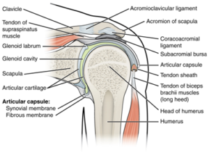
The shoulder joint (also known as the glenohumeral joint) is a ball and socket type joint, similar to that of the hip, that consists of three primary bones: the humerus (upper arm bone), the scapula (shoulder blade), and the clavicle (collarbone).

The head of the humerus fits into the shallow socket of the scapula, forming the glenohumeral joint. This is then held into the socket via active and passive structures. The active structures being the Rotator Cuff and the passive structures are the ligaments and joint capsule of the shoulder.

These key structures are involved in the shoulder’s mobility and overall function, and it is damage, overuse or underuse of one of these structures that can cause acute or chronic shoulder pain and dysfunction.

First let’s take a look at the active stabilizers of the shoulder; The Rotator Cuff. The rotator cuff is a group of four muscles that act as a suction cup around the head of the humerus. These muscles are constantly working together as one unit throughout normal shoulder movement to stabilize the humeral head in the socket and produce smooth movement. These muscles include the supraspinatus, infraspinatus, teres minor, and subscapularis.
When movement is produced friction occurs between the tendons and bones. In order to reduce this friction we have structures called Bursa which are filled with fluid and lubricate and reduce overall friction between the moving tendons of the shoulder.

Now let’s take a look at the passive stabilizers of the shoulder, starting with the Glenoid Labrum. The labrum is a rim of cartilage that wraps around the circumference of the glenoid fossa (the socket) in order to deepen the socket and provide a little more support for the moving humeral head.
This is then supported with the joint capsule, the joint capsule is an envelope surrounding the joint filled with synovial fluid which is required for smooth movement.

The bones which make up the shoulder joint are connected via ligaments. Which is a dense connective tissue that connects bone to bone – providing stability and support between the main bones of the shoulder.

Now we’ve taken a look at the anatomy of the shoulder, we can understand that there are many different structures around the shoulder which can become dysfunctional and as a result throughout the normal movement of the shoulder and cause pain.

Rotator Cuff Injuries:

Rotator cuff injuries come in a variety of forms, from acute traumatic injuries such as tears or strain ,of the rotator cuff muscles, or can be more of a chronic condition from overuse and degeneration and resulting in ongoing pain and limited mobility.

Frozen Shoulder (Adhesive Capsulitis): A condition characterized by stiffness and restricted range of motion due to inflammation and thickening of the shoulder capsule. This can be debilitating and happen post surgery, after a traumatic incident or be idiopathic (of unknown origin).

Bursitis: Inflammation of the bursa can occur due to repetitive overhead motions or direct trauma, causing pain and swelling in different areas of the shoulder joint. Typically this is accompanied by night pain, a burning sensation and weakness in the movements of the shoulder.

Tendon Related Pain: Tendon related pain is usually due to an overuse or an overloading of the muscles of the shoulder which can cause inflammation around the tendon and microdamage to the tendon itself.

Arthritis is a painful condition that occurs over time with natural degenerative changes to the joint. Arthritis is a normal process of aging however can be painful and debilitating if not treated appropriately.

Neck related shoulder pain: In some cases, shoulder pain can often be referred from the joints, muscles or nerve roots that stem from the cervical spine (neck). This can lead people to believe that shoulder pain is coming from the structures in the shoulder, however it can often be referring from a different location, making it difficult to treat and diagnose without an experienced Physiotherapist.

Shoulder Instability: Shoulder instability occurs due to the disengagement, inactivity or overactivity of the muscles of the shoulder, due to a shallow ball and socket joint as spoken about previously, due to a traumatic injury which has caused structural damage and can be a debilitating and frustrating injury which requires an accurate diagnosis and management plan to rehabilitate.

This is only scratching the surface when it comes to the shoulder joint, there is still so much we are learning through research and there is still so much unknown. The shoulder is a very complex joint which does not have a one size fits all solution. At Team Elite Health we have the skills necessary to accurately diagnose, manage and rehabilitate shoulder related pain and injuries to ensure that quality of life is restored and you are able to return back to full function of daily life, whatever that may be for you.

Hip and Groin Pain – Exploring FAI and Dysplasia. Two Common Causes

Hip Pain

Hip and groin pain can be debilitating, affecting one’s quality of life and mobility. Two very common conditions associated with this discomfort are Femoroacetabular Impingement (FAI) and Hip Dysplasia (DDH). In this comprehensive blog post, we will delve into these conditions, exploring their causes, symptoms, diagnosis, and potential treatment options to help you gain a better understanding of these two common causes of hip and groin pain.

First let’s take a deeper look into the hip joint itself in order to gain a better understanding of how it works, the structures surrounding it and possible causes for symptoms coming from the hip. Like we spoke about in the Shoulder Blog post, the hip is a ball and socket joint which is composed of the head of the femur (the ball) and the acetabulum of the pelvis (the socket).

Being a ball and socket joint, the hip joint moves in all planes of motion, which is initiated by the muscles surrounding the hip joint. Some examples of these muscles include the hip extensors, hip flexors, hip abductors, hip adductors and the hip internal and external rotators.

The passive structures of the hip include the ligaments surrounding the head of the femur and the femoral labrum, which like the shoulder joint, provides more depth to the ball and socket of the hip creating for more stability.

There are two structural causes for pain to occur in the hip joint; first lets take a look at Femoroacetabular Impingement, or FAI.

Femoroacetabular Impingement (FAI)

Femoroacetabular Impingement, commonly known as FAI, is a condition where there is abnormal friction between the hip’s ball and socket joint. This friction typically occurs when the hip’s ball (femoral head) or the socket (acetabulum) has an irregular shape, causing them to rub against each other during movement.

This is often caused throughout childhood development and is directly related to the activity that was completed during development of the bones. When in development, the body’s natural response to excessive loading is to throw more bone structure down in order to provide more support to the hip joint, however this can become problematic in the later stages of life.

There are three primary types of FAI:

  • Pincer Impingement: This occurs when the acetabulum (the socket) has an overgrowth of bone around the top of the femoral head, leading to a pinching effect during movement due to the bone pinching on the head of the femur.
  • Cam Impingement: In cam impingement, the femoral head is not perfectly round and has bumpy and protruding parts which affects the normal smooth movement of the ball in the socket and may lead to grinding against the acetabulum, or restricting the overall movement of the joint.
  • Combined Impingement: As the name suggests, this type combines both pincer and cam impingement, making it a more complex condition.

Symptoms of FAI typically include hip and groin pain, limited range of motion, and discomfort during specific activities. Diagnosis involves a thorough examination of medical history, physical assessment, and imaging studies like X-rays and MRI scans to identify impingement types and assess joint damage. However it is important to note that most people scanned that have a pincer or cam morphology have no symptoms (asymptomatic).

Treatment options for FAI range from conservative approaches, such as physiotherapy and anti-inflammatory medications, to more invasive methods like arthroscopic surgery.

Generally when first diagnosed, by improving the strength and range of motion of the hip joint symptoms will often decrease depending on the severity. Where symptoms are not improving or conservative management isn’t working, arthroscopic surgery may be an option or total joint replacement can also be considered. However most patients get significant outcomes with physiotherapy and have no need for surgery

The goal is to alleviate symptoms, improve hip function, and, when necessary, reshape the joint to mitigate impingement. Timely intervention is crucial to managing FAI effectively and restoring patients to an active and pain-free lifestyle.

Hip Dysplasia

Hip dysplasia is a developmental disorder affecting the hip joint’s structure, where the hip socket (acetabulum) is abnormally shallow, leading to instability. Generally this is diagnosed in babies at birth, but can also be missed and only present symptoms later in life.

This condition often manifests as a misalignment and a shallow surface between the femoral head (ball) and acetabulum, causing increased friction, movement and potential joint damage if left untreated.  This lack of congruence can result in joint instability slipping and feelings of clicking, popping and a feeling of instability throughout the hip joint. While some individuals with hip dysplasia may remain asymptomatic, others may experience pain, instability, and reduced range of motion.

Diagnosis involves a combination of physical examinations and imaging studies like X-rays and MRI scans to assess hip joint stability and identify the degree of dysplasia. Symptoms of hip dysplasia can vary, including hip or groin pain, a sense of joint instability, and limitations in daily activities.

Treatment options for hip dysplasia depend on the severity of the condition. Conservative approaches may include physiotherapy and lifestyle modifications, while more severe cases may require surgical interventions like pelvic or periacetabular osteotomy to improve joint stability and function. Early detection and appropriate management are crucial in addressing hip dysplasia, aiming to relieve symptoms and enhance the overall quality of life for individuals affected by this condition.

Hip and groin pain can have a significant impact on one’s daily life, but with a better understanding of conditions like FAI and hip dysplasia, individuals can seek early diagnosis and appropriate treatment. Whether it’s through conservative management, physiotherapy and strengthening, or surgical interventions, there are options available to alleviate pain and improve hip joint function.

If you’re experiencing hip and groin pain, it’s essential to consult with a healthcare professional for an accurate diagnosis and personalized treatment plan. Remember that addressing these issues promptly can help you maintain an active and pain-free lifestyle and reduce the risk of complications later on in life.

The Benefits of Sports and Recreational Activities for Adolescent Development

Adolescent

It is well known across the globe that there are a multitude of benefits of living an active lifestyle, in today’s blog we will be highlighting the benefits of sports, recreational activities and living a healthy lifestyle particularly for adolescents and their development into adulthood.

Adolescence, otherwise known as the pre adult phase or childhood is a crucial period of physical, emotional, and psychological growth in an individual’s life. Its a time where navigation is made from the path of childhood to adulthood and the choices that are made throughout this period can significantly impact the growth and development of their lives.

Engaging in physical activity such as sports and recreational activity is a powerful and beneficial way for adolescents to shape their development on a multitude of levels.

In this comprehensive blog post, we will explore the myriad benefits of sports and recreational activities for adolescents, highlighting how participation in these activities can shape their physical, social, emotional, and cognitive development.

The first and foremost and primarily the most rewarding aspect of engaging in physical activity is the direct influence on Physical Health and Fitness. Now we can break this up into a few different aspects of what Physical Health and Fitness really means.

  • Physical Fitness: Sports encourage regular exercise, helping adolescents maintain healthy body weight, develop strong muscles and bones, and improve cardiovascular health.
  • Weight-bearing and resistance exercise play vital roles in developing muscle mass and attaining peak bone density in early adulthood, important for musculoskeletal health in later life.
  • Improve performance: The increases in strength, muscle mass and bone density will help individuals excel at their chosen sports and be able to reach their peak performance.
  • Reduce Injury Risk: By improving function of the muscles, bones and ligaments, this will ultimately allow the adolescent to prevent injury from occurring due to meeting the specific demands of sport.
  • Motor Skills: Adolescents develop fine and gross motor skills through activities like swimming, dancing, and team sports. This enhances coordination, balance, and agility.
  • Resistance to Disease: Physical activity boosts the immune system, reducing the risk of various illnesses and chronic conditions, including obesity, diabetes, and heart disease.
  • Lifelong Habits: Engaging in sports and recreational activities during adolescence instills a habit of physical activity that can be carried into adulthood.

The second most rewarding aspect of engaging in Physical activity / exercise is the emotional and psychological and social development that is attained with organized sport / physical activity.

Social Development:

Participating in group activities fosters social development in adolescents, teaching them essential life skills and interpersonal relationships:

  • Teamwork: Team sports like footy, soccer, basketball, and netball teach adolescents how to collaborate, communicate, and work collectively towards a common goal.
  • Leadership: Some adolescents assume leadership roles within their sports teams, honing their leadership skills and learning to motivate and inspire others.
  • Communication: Adolescents learn to express themselves effectively, whether it’s on the field, court, or in the change rooms.
  • Conflict Resolution: Sports often involve disputes and disagreements, offering valuable opportunities for adolescents to learn conflict resolution and negotiation skills.
  • Friendship and Belonging: Adolescents build friendships with teammates and experience a sense of belonging and camaraderie that contributes to their self-esteem and social well-being.

Emotional Development:

Sports and recreational activities provide adolescents with a safe space to explore and manage their emotions – often using it as a release mechanism.

  • Self-Confidence: Achieving personal and team goals in sports boosts self-esteem and self-confidence.
  • Stress Reduction: Physical activity releases endorphins, which reduce stress and improve mood, helping adolescents cope with the emotional challenges of adolescence.
  • Emotional Regulation: Adolescents learn to manage disappointment, anger, and anxiety in the context of sports, valuable skills they can apply to other areas of life.
  • Goal Setting: Setting and achieving sports-related goals can help adolescents develop a sense of purpose and direction.
  • Understanding winning and losing: Normalises both winning and losing in life which prepares a young adolescent to adult life.

Social Development:

Participating in group activities fosters social development in adolescents, teaching them essential life skills and interpersonal relationships from working in a team environment.

  • Teamwork: Team sports like footy, soccer, basketball, and volleyball and netball teach adolescents how to collaborate, communicate, and work collectively towards a common goal.
  • Leadership: Some adolescents assume leadership roles within their sports teams, honing their leadership skills and learning to motivate and inspire others.
  • Communication: Adolescents learn to express themselves effectively, whether it’s on the field or in preparation for an event or game.
  • Conflict Resolution: Sports often involve disputes and disagreements, offering valuable opportunities for adolescents to learn conflict resolution and negotiation skills.
  • Friendship and Belonging: Adolescents build friendships with teammates and experience a sense of belonging and camaraderie that contributes to their self-esteem and social well-being.

Emotional Development:

Sports and recreational activities provide adolescents with a safe space to explore and manage their emotions:

  • Self-Confidence: Achieving personal and team goals in sports boosts self-esteem and self-confidence.
  • Stress Reduction: Physical activity releases endorphins, which reduce stress and improve mood, helping adolescents cope with the emotional challenges of adolescence.
  • Emotional Regulation: Adolescents learn to manage disappointment, anger, and anxiety in the context of sports, valuable skills they can apply to other areas of life.
  • Goal Setting: Setting and achieving sports-related goals can help adolescents develop a sense of purpose and direction.

Cognitive Development:

Engaging in sports and recreational activities can positively impact cognitive development in adolescents:

  • Problem Solving: Team sports often require quick thinking and adaptability, fostering problem-solving skills.
  • Concentration: Sports demand focus and concentration, which can translate to better academic performance.
  • Discipline and Time Management: Balancing school, sports, and personal life teaches adolescents valuable time management and discipline.
  • Goal Orientation: Learning to set and achieve goals in sports can transfer to academic and career aspirations.

Character Development:

Participation in sports and recreational activities helps shape adolescents’ character in numerous ways:

  • Perseverance: Adolescents learn to persevere through challenging practices and games, developing resilience.
  • Sportsmanship: Adolescents learn the importance of fair play, integrity, and respect for opponents and officials.
  • Accountability: Being part of a team teaches adolescents accountability for their actions and responsibilities.
  • Work Ethic: Commitment to training and practice instills a strong work ethic.

Healthy Lifestyle Choices:

Adolescents involved in sports and recreational activities are more likely to make healthy lifestyle choices:

  • Nutrition: Sports often come with nutritional education, promoting healthy eating habits which fuel the athlete and promote growth and athletic development.
  • Avoiding Risky Behaviors: Adolescents engaged in sports are less likely to engage in risky behaviors such as underage drinking, and staying out of trouble.
  • Better Sleep: Regular physical activity can improve sleep patterns, ensuring adolescents get adequate rest which is crucial to development academically and physically.

Participating in sports and recreational activities during adolescence offers a wide range of benefits that extend beyond physical health. These activities promote social, emotional, cognitive, and character development, equipping adolescents with valuable life skills and experiences that will serve them well throughout their lives. Encouraging adolescents to engage in sports and recreational activities is an investment in their holistic development, contributing to healthier, more confident, and well-rounded individuals ready to face the challenges of adulthood.

Understanding the Science behind Hot and Cold Therapy

Hot and cold bath

Hot and cold therapy has been used throughout the sporting world as a widely recognised tool for managing muscle soreness, inflammation and overall recovery post exercise and sport activity. Hot and cold therapy utilize the body’s natural response of thermoregulation (the ability to maintain a constant body temperature) by creating a vascular response to improve overall healing and promote optimal muscle conditions and ultimately function.

Hot and cold therapy seems like such a simple concept, however, their effectiveness stems from complex physiological functions that harness the power of the body’s response to outside temperature changes is how the process works. Although used widely across the sporting population, the science behind how it works is not vastly understood. The purpose of this blog is to help athletes, practitioners and patients understand the science behind the therapy in order to understand and unlock the potential for maximizing recovery and ultimately improving performance in sport and everyday life.

Hot Therapy (Thermotherapy)

As the name states, hot therapy or thermotherapy is the process of applying heat to affected areas in various modalities such as full body immersion, heat pads, warm compresses or more recently with technology such as infrared sauna. The process and physiological steps are the same for all of the thermotherapy modalities; Vasodilation.

Vasodilation translates to the dilation of blood vessels within the body. When blood vessels dilate, this means that they open and allow for greater blood flow throughout the body including but not limited to muscles around the body. Increases in muscle blood flow aid in the recovery from intense, muscle-damaging exercise through several different possible mechanisms:

First, the delivery of substrates in the blood such as glucose and essential amino acids that are required for refueling and tissue rebuilding is enhanced which ultimately will lead to an increased rate of tissue healing.

Second, the removal of by-products of exercise such as hydrogen ions (responsible for soreness in muscles post exercise) as well as other substances that can cause a delayed healing response is targeted and removed at a faster rate due to the increase of blood flow to the affected areas. (Kim, Monroe, Gavin, & Roseguini, 2020)

Thermotherapy also helps with the feeling of tightness throughout the body which is usually felt post exercise. This occurs when the muscle fibers shorten and stay in a shortened position post exercise. Heat therapy activates receptors within the brain (TRPV1) receptors which help to regulate pathways through the nervous system which affect muscle tonicity (tightness) and relax muscles. (Malanga, Yan, & Stark, 2014) Heat also helps to cause an increase in elasticity of connective tissue called fascia, which ultimately will help to reduce spasms, stiffness, musculoskeletal pain and increase muscle flexibility.

Cold Therapy (Cryotherapy)

Unlike Thermotherapy, Cold therapy or Cryotherapy is application of something cold, whether that is submersion in cold water, application of ice or an ice pack to a certain area, use of a game ready machine or whole body ice therapy in an ice bath.

The primary mechanism that occurs when cryotherapy is applied to the body is called Vasoconstriction or the narrowing of the blood vessels within the body. Cold causes the blood vessels to constrict, restricting blood flow to the region. This can help alleviate swelling and inflammation which are often common after exercise and activity which causes micro damage to the muscle tissue.

When we move and when we exercise we are breaking down stored energy in the muscle. As a result of this process, the body produces by products, think of it like the exhaust of your car when you drive. The natural process of elimination of these by-products is via 2 mechanisms, it is transported via blood flow and recirculated around the body or it is used again in the muscle as a source of energy. However, this process doesn’t always go smoothly and often these by-products are left behind in the muscles and get stuck. This combined with the microtrauma that occurs with exercise creates a feeling of soreness.

This is when we feel delayed onset of muscle soreness or DOMS (delayed onset muscle soreness). The soreness we feel after exercise. The combination of microtrauma as well as pooling of blood in the vessels creates a feeling of soreness and stiffness post exercise.

Vasoconstriction helps with constricting the blood vessels within the muscle, this mechanism helps to squeeze any pooling blood that is left over or stuck within the muscle and flushes it out of the affected tissue.

Cold temperatures also reduce blood flow to the affected area via vasoconstriction. This has been thought to reduce swelling and tissue breakdown after an injury or after procedures such as surgery.

Combination Therapy

An increasingly more popular form of recovery post exercise is known as combination therapy, combining the use of thermotherapy and cryotherapy together in contrast with each other. This method of therapy utilizes the two modalities we mentioned earlier, vasoconstriction and vasodilation. Alternating between these two methods of recovery induces a pumping effect within the vascular system.

Ultimately this pumping effect helps to both squeeze left over waste products of exercise and then allow for an influx of fresh blood flow to the muscles which results in increased oxygen, increased essential amino acids and removal of waste products and swelling which will overall improve lower limb function, reduce soreness within the muscles and promote a quicker recovery. (Shadgan, Pakravan, Hoens, & Reid, 2018).

By repeating these two methods within a 20-30 minute session the constant changing of temperature and vascular size has been shown to improve recovery and reduce the overall DOMS felt post exercise which can get an athlete ready for performance quicker, reduce overall fatigue and therefore injury risk and allow for harder training at their next session.

Kim, K., Monroe, J. C., Gavin, T. P., & Roseguini, B. T. (2020). Local heat therapy to accelerate recovery after exercise-induced muscle damage. Exercise and Sport Sciences Reviews, 48(4), 163–169. doi:10.1249/jes.0000000000000230

Malanga, G. A., Yan, N., & Stark, J. (2014). Mechanisms and efficacy of heat and cold therapies for Musculoskeletal injury. Postgraduate Medicine, 127(1), 57–65. doi:10.1080/00325481.2015.992719

Shadgan, B., Pakravan, A. H., Hoens, A., & Reid, W. D. (2018). Contrast baths, intramuscular hemodynamics, and oxygenation as monitored by near-infrared spectroscopy. Journal of Athletic Training, 53(8), 782–787. doi:10.4085/1062-6050-127-17

Apophysitis – The Growing Disease

Apophysitis

What is it? Why does it Occur?

Apophysitis, also known as an apophyseal injury, is a condition that primarily affects children and adolescents, especially those who are involved in sports that involve repetitive movements, are high impact (running, jumping, landing, cutting, pivoting). Apophysitis is primarily seen in the younger population and is caused by overuse, creating inflammation and therefore pain at the site of attachment of tendons on areas of bone known as the growth plates or physis.
The reason this injury primarily occurs in children and adolescents is due to the nature of the bone, tendon and cartilage.
Muscles attach to tendons which then attach onto cartilage and bones. In certain areas of the body these tendons attach onto growth plates which are skeletally immature as they allow for growth of the bones.
This creates a weaker attachment site which means the tendon and bone complex is unable to resist stress (particularly shear and tension forces) compared to the cartilage and bone in adults.


What are the risk factors?

The most common risk factors for apophysitis are periods of rapid growth. During these periods there is a mismatch of the rate of growth between bone and muscle. Typically bone will start to grow first, followed by muscle growth. This mismatch creates tension and therefore traction on the soft attachment site which leads to pain, inflammation and swelling.
Combine the growing with sports, usually multiple sports nearly every day. Exercising and in particular running sprinting jumping will cause an increase in traction on the attachment site of the tendon, further causing pain swelling and inflammation.

What are the Symptoms?

Symptoms of apophysitis usually include;

  • Pain, tenderness and often swelling at the affected area
  • Pain worsens with physical activity
  • Reduced range of motion and discomfort with movement at the affected muscle/tendon junction

Common Types

As there are multiple growth plates located across the body, the more common types are seen in the lower limb or legs in the young athlete, however, it is possible to have apophysitis in the upper limb.

Calcaneal Apophysitis (Sever’s Disease)

Calcaneal apophysitis, better known as Sever’s Disease, is considered one of the most common diagnoses of posterior heel pain in rapidly growing, physically active children aged between 8 and 15 years (Volpe et al., 2009; Howard, 2014)

Sever’s Disease is described as an ‘overuse injury’ with a gradual onset of pain with no specific traumatic event in the physically active growing children population (Ramponi et al., 2019)
Sever’s disease can present unilaterally or bilaterally, with an incidence of 60% of cases presenting bilaterally (Smith et al., 2021)

Risk factors for Sever’s Disease

  • Considered an ‘overuse’ injury, an increase in sports activity and/or training volumes (particularly sports requiring repetitive running and jumping – Smith & Varacallo, 2022)
  • Achilles tendon tightness
  • Poor ankle dorsiflexion strength
  • Inadequate shoes (including worn-out athletic shoes)
  • Environmental (running on harder surfaces etc).
  • Biomechanical factors that contribute to poor shock absorption (foot shape, proximal limb weakness etc)– James et al., 2016
  • Obesity
  • Gender (boys 3:1 ratio to girls)

Tibial Tuberosity Apophysitis (Osgood Schlatters Disease)

Considered one of the most common overuse injuries of the lower limb causing anterior knee pain in the young, skeletally immature athletic population – with basketball, volleyball, sprinters, gymnastics and football the most common sports associated with the condition (Smith & Varacallo, 2022; Corbi et al., 2022)
Osgood-Schlatters is atraumatic with a, gradual onset of pain that is increased with physical activity (Smith & Varacallo, 2022)

Risk factors for Osgood Schlatters Disease

  • Gender (males have a higher likelihood of developing OSD)
  • Adolescents aged 8-15 years
  • A recent, rapid increase in growth
  • Increased levels of physical activity
  • Hamstring and Quadriceps tightness (particularly rectus femoris)
  • As previously mentioned, adolescents participating in sports that require repetitive jumping and/or running (basketball, volleyball, athletics, gymnastics and football)
  • Some studies have identified that reduced elasticity of the patella tendon can be associated with a greater risk of OSD (Enomoto et al., 2020)


Ischial Tuberosity / Hamstring Apophysitis

Like Sever’s and Osgood-Schlatter’s, hamstring apophysitis is an ‘overuse’ injury which results in inflammation of the apophysis that undergoes ossification to form the ischial tuberosity during adolescence.
The location of pain is situated at the proximal hamstring attachment to the ischial tuberosity apophysis and may produce pain in the gluteal region when performing physical activity (including sports with high physical plyometric demands such as running, football, rugby and tennis)

Risk factors for Hamstring Apophysitis

  • Physically active young adolescents
  • Increased training volumes (pre-season, competitions)
  • Rapid growth spurt (with bones growing faster than muscle tissues and reducing flexibility)
  • Linking to above, reduced flexibility of the hamstring muscles (biceps femoris, semitendinosus and semimembranosus)
  • Type of sport (similar to Sever’s and Osgood-Schlatters with the young athletic population performing repetitive jumping and running at a higher risk of developing apophysitis)
  • Hamstring to quadriceps strength ratio

So how do we prevent or treat Apophysitis?

Apophysitis can be debilitating in season and cause lots of pain and dysfunction in the active adolescent.
Prevention is always better than cure and there are many ways to prevent this common injury occurring.

Physiotherapists are highly trained in all of the above prevention strategies and can be a crucial part of an adolescents active lifestyle. By improving overall lower limb strength with a specific tailored program has shown to have a significant reduction in pain, inflammation and swelling and decrease the overall prevalence of this overuse injury.
Treatment for Apophysitis varies from individual to individual however ultimately starts with a basic strength and biomechanical assessment to see whether any patterns in movement can be contributing factors to the progression of the injury. From this assessment a specific strengthening and loading programme can be created which is tailored to the individual. Without changing biomechanics or altering the amount of strength in certain muscle groups, the problem is not being addressed and will continue to cause issues as the adolescent goes through growth spurts.

References

Smith JM, Varacallo M. Sever Disease. [Updated 2021 Jul 18]. In: StatPearls [Internet]. Treasure Island (FL): StatPearls Publishing; 2021

Volpe R. Keys to diagnosing and treating calcaneal apophysitis. Podiatry Today. 2009;22(11):60-66.

Lucenti L, Sapienza M, Caldaci A, Cristo C, Testa G, Pavone V. The Etiology and Risk Factors of Osgood-Schlatter Disease: A Systematic Review. Children (Basel). 2022 Jun 2;9(6):826. doi: 10.3390/children9060826. PMID: 35740763; PMCID: PMC9222097.

Vaishya R, Azizi AT, Agarwal AK, Vijay V. Apophysitis of the Tibial Tuberosity (Osgood-Schlatter Disease): A Review. Cureus. 2016 Sep 13;8(9):e780. doi: 10.7759/cureus.780. PMID: 27752406; PMCID: PMC5063719

Kujala UM, Orava S, Karpakka J, Leppävuori J, Mattila K. Ischial tuberosity apophysitis
and avulsion among athletes. Int J Sports Med. 1997 Feb;18(2):149-55. doi: 10.1055/s-2007-972611. PMID: 9081273.

5 Common Sporting Injuries of the Lower Body and How to Prevent Them

Performance Testing

Ankle sprain

Ankle sprains, or “rolled ankles” are very common injuries in football, soccer, basketball and netball. Sprains occur when the ligaments of the ankle are put under tension with running, change or direction or jumping and landing. If the force through these ligaments is too great, they can be stretched or torn.

Strength training of the lower limb muscles, proprioception or balance training and a focus on jumping and landing mechanics are all helpful in preventing an initial ankle sprain. In addition these are also extremely important in a rehabilitation program after an ankle sprain. A prehabilitation program guided by a Team Elite Physiotherapist would be a great way to build a strong and stable ankle. In addition to this, there is also evidence in the use of ankle tape or bracing to prevent recurring ankle ligament injuries (Vuurberg et al., 2018) and are worth discussing with your treating health practitioner.

Knee ligament injuries

There are four main ligaments that support and stabilize the knee joint, which are: the Anterior and Posterior Cruciate Ligaments (ACL and PCL) which support the knee from front to back, and the Medial and Lateral Collateral Ligament (MCL and LCL) which support the knee side to side. Injury to any of these can affect the stability of the knee and cause buckling

Quality warm ups focusing on activation and movement patterns, attention to running mechanics and change of direction practice focusing on knee and hip positioning and having high levels of strength in the lower limbs will all help at preventing these injuries. Our physiotherapy department at Team Elite Health are very well educated in the preparation and rehabilitation of knee ligament injuries and can assist anyone needing help or guidance with a strength program or biomechanical assessment. In addition to helping streamline surgical appointments and post surgical recovery with our state of the art facilities.

Plantar Fasciitis

The plantar fascia is the broad band of connective tissue that connects the underside of the heel to the toes, holding up the arch of the foot. It is important in providing correct foot posture when pushing off from the ground while walking, running and jumping. Overuse of the plantar fascia can lead to a painful arch or heel when playing sport and even difficulty walking.

Your choice of footwear is a key factor in the prevention and management of plantar fasciitis. Shoes that are either too old and worn out, or shoes that aren’t the right support for your foot arch are both contributors to an overloaded plantar fascia. Combining the right shoes with a smart training plan are simple ways to reduce the risk of becoming sore.

Muscle strains

Soft tissue injuries are the most common injuries in sports. Of those, muscle strains represent a large number of these, with the hamstring muscle the most commonly injured in Australian Rules Football. Injuries to the hamstring often occur when running at high speed, kicking or changing direction quickly. They are usually easy to spot, with a sudden sharp pain or pull described as a “ping”, loss of range of motion, loss of power and pain walking and running.

While some risk factors like age and previous injury can’t be avoided, some can be mitigated. Previous hamstring injury is a significant risk factor for future injuries. Ensuring a complete and robust rehabilitation plan under the guidance of a physiotherapist will help to restore the muscles function to what it was prior to the injury and often even make it stronger and more resilient to an injury. Even without a previous hamstring injury, working closely with your physiotherapist or strength and condition coach to maximize your strength and high speed running ability can be beneficial and reduce your risk of hamstring strain.

Shin Splints

Shin splints is a commonly used term given to pain experienced along the front and inside of the shin, often affecting running based athletes. It is important to note that shin splints is not an actual diagnosis, but rather an umbrella term used to describe the location of symptoms that includes numerous causes. There two most common causes of shin pain in this context are Medial Tibial Stress Syndrome (MTSS) and stress fractures of the tibia or shin bone.

While the conditions differ in their presentation and treatment requirements, they do share a few common risk factors or potential causes. High training loads or large fluctuations in activity, poor lower limb biomechanics whilst running or jumping, incorrect or old footwear and being overweight can all contribute to the development of MTSS or stress fracture. Starting exercise slowly and reducing the overall volume of your training to be pain free, wearing the correct footwear, reviewing your running technique and commencing a lower limb strengthening program with a suitably qualified professional and reducing body weight are all modifiable factors that will help to reduced the risk of injury (Hamstra-Wright, Bliven & Bay; 2015).

5 common sporting injuries of the upper body and how to prevent them

sporting injuries

Australians are known for their love of sport, with approximately 41% of us participating in sport related activities at least once per week (Australian Sports Commission, 2022). Regular sporting involvement can help both physical and mental health, improve sleep quality, help to develop discipline and routine and help to form important social connections. However, playing sports is not without its downsides, and getting injured is one of them. In this post, we will talk through 5 common upper body injuries.

Concussion

Concussions are a type of head injury that occur with a direct knock or blow to the head, and are considered mild traumatic brain injuries that result in compromised brain function. Common symptoms include headaches, confusion, dizziness, memory loss, loss of consciousness, nausea and vomiting, mood swings and fatigue. Early identification and appropriate management of these injuries are critical to the long term health outcomes of the injured player.

While not all concussions can be avoided, there are a number of suggested ways to reduce the frequency and severity (Waltzman & Sarmiento, 2019). Firstly, educating all players, staff and parents as to the signs and symptoms of a concussion allows for the early identification, which will lead to better outcomes for the injured athlete. Secondly, using protective equipment that is in good condition and that meets Australian safety standards can reduce the impact forces associated with the collision. Lastly, strengthening of the neck muscles has been shown to help absorb some of the forces associated with concussions.

Dislocations

Dislocated joints are another common injury presenting to physiotherapy clinics, often sustained while playing ball or contact sports. Dislocated shoulders, fingers and patellas (knee caps) are often seen across a range of sports.

Joints, or the meeting of two bones, allow for smooth pain free motion between the segments which allow us to move. Joints are supported by ligaments, capsules that surround the joint and even tendons or muscles passing along the joint. Under normal pressures and loads, these structures are able to stabilize the joint and keep it together. Dislocations occur when the bones are forced apart by high external forces, such as a collison or tackle during sport, a fall, or a sudden change of direction. If the joint moves only a little and is not completely separated, it is called a subluxation. If however the joint surfaces are completely disengaged, this is known as a dislocation. In both cases, there may be damage to the ligaments and other structures that support the joint.

Dislocated shoulders and fingers seem to be the prevailing dislocations seen by physiotherapists in our clinic. Whilst some joint injuries are accidental and unavoidable, there are known risk factors that we should be mindful of, including weakness in the surrounding muscles, stretched ligaments from previous dislocations, joint hypermobility, connective tissue disorders such as Ehlers-Danlos syndrome or Marfan syndrome. In all of these examples, a thorough strength program targeting specific stabilizing muscles has been proven to be effective at reducing the risk of dislocations. Furthermore, the use of tape whilst playing sport can help to support the joint and reduce the chance of a dislocation (Turgut et al., 2021).

Shoulder Impingement or Pinching Shoulder Syndrome

The shoulder joint is one of the most mobile joints in the body, allowing for very large ranges of motion in multiple directions. Not surprisingly, this means that the shoulder is susceptible to being injured, often affecting overhead and throwing athletes. Shoulder impingement refers to the compression, irritation and inflammation of the structures that sit in the subacromial space of the shoulder. These structures become progressively more irritated and begin to cause pain, which also leads to a loss of shoulder movement overhead and sometimes pain at night when lying on that shoulder.

Common causes of shoulder impingement include poor strength of the shoulder rotator cuff muscles, particular shapes of the tip of the acromion bone and thickened or degenerative rotator cuff tendons; all leading to a narrowing of the subacromial space. Furthermore, poor mobility of the scapular itself can orient the shoulder downwardly, contributing to an apparent loss of subacromial space. . Short term relief may be possible with anti-inflammatory medication, however successful management depends on the accurate identification of the contributing factor and targeted exercise and strengthening to correct this (Consigliere et al., 2018).

Tennis Elbow

Lateral epicondylitis, or tennis elbow, is an injury that presents with pain on the outside of the elbow. It is most commonly an overuse injury of the muscles in the forearm that extend the wrist, but sometimes can be caused by an acute traumatic injury to the elbow. Whilst it is not an injury exclusive to the sport of tennis, a large number of cases are those that play tennis.

Like most overuse injuries, ensuring the correct load or amount of exercise is a key preventative strategy. Working closely with a coach to hone your stroke, getting the right grip size and string tension and not over training are easy ways to avoid becoming injured. Strengthening the entire upper limb, from grip strength all the way to shoulder stabilizers is must in order to stay on the court (Nabil et al., 2019).

Golfers Elbow

Like Tennis Elbow, Golfer’s Elbow is an overuse injury of the elbow where pain is typically seen on the inside of the elbow and forearm. Again, the condition is not only seen in those playing golf. Sports involving repeated throwing or gripping, and physical jobs like trades are common causes of Golfer’s Elbow.

Taking a period of time to rest from the activities that hurt, while ice and anti-inflammatory medications are of limited benefit. The most effective way to manage Golfer’s elbow is through physiotherapy, which may include things like dry needling, shock wave therapy, and strengthening of the upper limb and counterforce braces (Hoogvliet et al., 2013). Rarely do these elbow overuse injuries require surgery, so it is best to make an appointment with your Physiotherapist to assess and begin your rehabilitation.

Stay tuned for the second part to this post where we will look at common lower body injuries. 

Hoogvliet P, Randsdorp MS, Dingemanse R, Koes BW, Huisstede BM. Does effectiveness of exercise therapy and mobilisation techniques offer guidance for the treatment of lateral and medial epicondylitis? A systematic review. Br J Sports Med. 2013 Nov;47(17):1112-9 

Consigliere P, Haddo O, Levy O, Sforza G. Subacromial impingement syndrome: management challenges. Orthop Res Rev. 2018;10:83-91.

The Importance of Physiotherapy in Sports Performance and Injury Prevention

Physiotherapy

Physiotherapy in Sports Performance

There is a common belief that higher training loads are associated with higher injury rates, however there is evidence which supports physiotherapy and strength training as a protective factor against injury and improving performance.

Physiotherapy is an essential discipline in which both professional and amateur athletes can rely on to give expert advice to enhance their physical competence, prevent injuries and thus improve overall performance. Physiotherapy plays a role in injury prevention in sports, through carefully tailored strengthening programs which address deficits in strength, thus improving an athlete’s performance in their specific sport. Physiotherapists help with the promotion of safe physical activity participation, provision of advice and adaptation of rehabilitation and training interventions for the purposes of preventing injury.

Common causes of sporting injuries which may cause athletes to miss time from their sport include overuse, poor biomechanics and reduced conditioning.

Overuse injuries

Physiotherapy in Sports Performance

Overuse injuries such as stress fractures and tendinopathy are highly prevalent and contribute to a high amount of time spent away from sporting activities. Unlike acute injuries – which are caused through sudden trauma, overuse injuries develop gradually over time due to repeated stress on tissues such as tendons, muscles, ligaments and bones (Launay, 2015). This occurs due to the body’s inability to repair and adapt to stressors placed on such tissues, thus leading to tissue damage, inflammation and associated pain. Physiotherapy is a necessary tool in avoiding overuse injuries as areas of weakness can be addressed and will incorporate load management techniques.

Tendon related injuries including quadriceps tendinopathy are associated with overuse, typically from repeated jumping, kicking sports, where the quadriceps cannot withstand load, leading for the tendon to become inflamed. Participating in early intervention from a physiotherapist helps reduce time away from sport. This is due to being provided with educated advice of load management techniques, icing and appropriate injury specific exercises to help increase strength and thus return the patient to play.

Lumbar or Back stress fractures are common overuse injuries in adolescents who participate in high levels of sport. Through having early intervention with carefully tailored exercise programs, patients are able to reduce inflammation from stress reactions, thus allowing the athlete to progress through rehabilitation and return to sport in a timely manner.

Poor biomechanics

Biomechanics is the study of how forces and movement interact through the body. When an individual has sub-optimal biomechanics, this means that their body is not moving efficiently and transferring force effectively. This may be due to a variety of factors such as muscle imbalances, reduced joint range of motion, poor technique or postural issues. By having poor biomechanics an individual will have increased risk of injury due to potential increase of forces through certain muscles/joints, poor compensation patterns and decreased energy efficiency due to excessive energy expenditure to perform movements (Hewett & Bates, 2017). These factors will also negatively affect performance in sport.

Athletes who run with poor knee drive, poor hip extension, increased hip adduction and increased extension or rotation through the trunk are not moving their body effectively thus reducing an individual’s ability to perform at their peak. Having physiotherapy input on biomechanics allows for conscious incompetence and then conscious competence. Conscious incompetence is the process of an individual being consciously aware of how to improve technique thus improving efficiency. Physiotherapists can highlight these issues and thus begin steps to improve performance. Over time individuals become consciously competent due to repetition of particular drills to improve efficiency, and eventually become unconsciously competent.

By incorporating physiotherapy into an athletes routine, biomechanical patterns can be positively changed through gait retraining, improve strength with a tailored strengthening routine and thus improve overall energy expenditure as the athlete moves more efficiently. Within the adolescent population biomechanical retraining is imperative for individuals who have suffered from conditions such as lumbar stress fractures, in order to reduce their risk of re-injuring of the same or new areas of the spine if the patient is running in a way which places extra stress in injury prone areas.

Reduced conditioning

Reduced conditioning refers to an individual’s decline in physical fitness which occurs due to inadequate training levels. Having reduced conditioning levels negatively impacts one’s performance, as it may cause a reduction in aerobic capacity and poor muscle strength. Having deficiencies in these areas will negatively influence one’s performance as they may not be able to effectively meet the demands of their sport. If an individual were to participate in a sport which exceeded their current conditioning level they are at risk of injury. Through participating in physiotherapy, individuals will be able to optimise performance through tailored strength training which takes into account their selected sport and own capabilities.

In the adolescent population we see reduced conditioning during time periods such as summer holidays as often this is where sports ceases for approximately two months. When this decline in activity levels occurs, the individual may be at risk of injuries such as stress fractures, apophysitis or tendinopathies, as once they return to sport their load exceeds their new reduced muscular capabilities.

It is imperative that after periods of reduced conditioning that individuals find a happy medium between overtraining and under training. Over training occurs when a person’s energy expenditure exceeds their body’s ability to recover from strenuous exercise. Under training occurs when there is a decline or stagnation of performance due to insufficient training frequency, volume, or intensity. With guidance of a physiotherapist an adequate preseason or in season program can be provided to reduce risk of deconditioning. Alternatively, a physiotherapist can help guide return to sport after a prolonged rest period to reduce the risk of over-training, thus reducing injury risk. By involving physiotherapy in an athlete’s routine such injuries can be avoided and thus increase the amount of time participating in sport related activities and athletic satisfaction. By reducing the risk of injury, focus can then be on improving sporting performance.

Launay, F. (2015). Sports-related overuse injuries in children. Orthopaedics & Traumatology: Surgery & Research, 101(1). https://doi.org/10.1016/j.otsr.2014.06.030

Hewett, T. E., & Bates, N. A. (2017). Preventive Biomechanics: A paradigm shift with a translational approach to injury prevention. The American Journal of Sports Medicine, 45(11), 2654–2664. https://doi.org/10.1177/0363546516686080

Sasaki, K & Neptune, R (2005) Muscle mechanical work and elastic energy utilization during walking and running near the preferred gait transition speed